Πέμπτη 10 Δεκεμβρίου 2020

Efficacy and Safety of Pembrolizumab (MK-3475) Plus Lenvatinib (E7080/MK-7902) Plus Chemotherapy in Participants With Advanced/Metastatic Human Epidermal Growth Factor Receptor 2 (HER2) Negative Gastric/Gastroesophageal Junction (GEJ) Adenocarcinoma (MK-7902-015/E7080-G000-321/LEAP-015)

pay.thelady61 shared this article with you from Inoreader

ct.gov-nlm-nih-logo.png

Condition:   Advanced/Metastatic HER2 Negative Gastric/Gastroesophageal Junction Adenocarcinoma
Interventions:   Biological: Pembrolizumab;   Biological: Lenvatinib;   Drug: Oxaliplatin;   Drug: Capecitabine;   Drug: Leucovorin;   Drug: 5-FU
Sponsors:   Merck Sharp & Dohme Corp.;   Eisai Inc.
Not yet recruiting
View on the web

Pilot Study of a 1-Millimeter Resolution Clinical PET System in Head and Neck Cancer Imaging

pay.thelady61 shared this article with you from Inoreader

ct.gov-nlm-nih-logo.png

Condition:   Head and Neck Cancer
Intervention:   Device: 1-mm Resolution Clinical PET camera
Sponsors:   Stanford University;   Goldman Sachs Foundation;   Wallace H. Coulter Foundation;   Emerson Collective
Not yet recruiting
View on the web

Systemic Inflammation Response Index Is a Predictor of Poor Survival in Locally Advanced Nasopharyngeal Carcinoma: A Propensity Score Matching Study

pay.thelady61 shared this article with you from Inoreader

575417_Thumb_400.jpg

Introduction

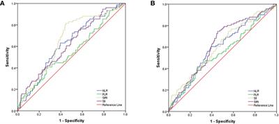
Nasopharyngeal carcinoma (NPC) is a common malignancy in China and known prognostic factors are limited. In this study, neutrophil-to-lymphocyte ratio (NLR), platelet-to-lymphocyte ratio (PLR), systemic immune inflammation index (SII), and systemic inflammation response index (SIRI) were evaluated as prognostic factors in locally advanced NPC patients.

Materials and Methods

NPC patients who received curative radiation or chemoradiation between January 2012 and December 2015 at the Second Xia ngya Hospital were retrospectively reviewed, and a total of 516 patients were shortlisted. After propensity score matching (PSM), 417 patients were eventually enrolled. Laboratory and clinical data were collected from the patients' records. Receiver operating characteristic curve analysis was used to determine the optimal cut-off value. Survival curves were analyzed using the Kaplan-Meier method. The Cox proportional hazard model was used to identify prognostic variables.

Results

After PSM, all basic characteristics between patients in the high SIRI group and low SIRI group were balanced except for sex (p=0.001) and clinical stage (p=0.036). Univariate analysis showed that NLR (p=0.001), PLR (p=0.008), SII (p=0.001), and SIRI (p<0.001) were prognostic factors for progression-free survival (PFS) and overall survival (OS). However, further multivariate Cox regression analysis showed that only SIRI was an independent predictor of PFS and OS (hazard ratio (HR):2.83; 95% confid ence interval (CI): 1.561-5.131; p=0.001, HR: 5.19; 95% CI: 2.588-10.406; p<0.001), respectively.

Conclusion

Our findings indicate that SIRI might be a promising predictive indicator of locally advanced NPC patients.

View on the web

Retention and survival rate of etanercept in psoriasis

pay.thelady61 shared this article with you from Inoreader

ABSTRACT

There have been a number of investigations of the efficacy and safety of etanercept. This study was performed to obtain long‐term drug survival data (i.e. time to drug discontinuation) for etanercept, and the reasons for its discontinuation. The study population consisted of patients with psoriatic arthritis and psoriasis followed up by our clinic, registered in the Turkish Psoriasis Registry (PSR‐TR) and treated with etanercept for at least 4 weeks between January 1, 2005, and January 31, 2020. The efficacy of etanercept was evaluated in terms of the Psoriasis Area and Severity Index (PASI) 75, PASI 90 and PASI 100 response rates at 12, 24, 36 and 48 weeks, and annually thereafter. The behaviours of the patients with respect to the use of etanercept, and the outcomes of those who continued to use it during the COVID‐19 pandemic, were also investigated.

This article is protected by copyright. All rights reserved.

View on the web

The Goldman‐Fox Syndrome: Treating and Preventing Green Pseudomonas Nails in the Era of COVID‐19

pay.thelady61 shared this article with you from Inoreader

Message:

Chloronychia: The Goldman-Fox Syndrome - Implications for Patients and Healthcare Workers
Robert A Schwartz 1, Nicole Reynoso-Vasquez 1, Rajendra Kapila 1
Affiliations collapse
Affiliation
1Department of Dermatology, Infectious Diseases, and Pathology, Rutgers New Jersey Medical School, Newark, New Jersey.
PMID: 32029931 PMCID: PMC6986112 DOI: 10.4103/ijd.IJD_277_19
Free PMC article
Abstract
Nail coloration has many causes and may reflect systemic disease. White nails (leukonychia) are common; rubronychia is rare, whereas green (chloronychia) is occasionally evident. Chloronychia, the Fox-Goldman syndrome, is caused by infection of an often damaged nail plate by Pseudomonas aeruginosa. P. aeruginosa is an opportunistic pathogen known for localized and systemic infections. It can spread cryptically in a variety of ways, whether from an infected nail to a wound either autologously or to a patient as a surgical site infection, and many represent a threat to elderly, neonatal, or immunocompromised patients who are at increased risk of disseminated pseudomonas infection. We will review the Goldman-Fox syndrome as an occupational disorder of homemakers, nurses, plumbers, and others often with wet hands. At a time when hand washing is being stressed, especially in healthcare settings, examination of nails should be emphasized too, recalling the possibility of surgical site infe ction even with a properly washed and gloved medical care provider. Pseudomonas may be a community-acquired infection or a hospital or medical care setting-acquired one, a difference with therapeutic implications. Since healthcare workers represent a threat of nosocomial infections, possible guidelines are suggested.

Keywords: Chloronychia; chromonychia; green; nails; pseudomonas.

Copyright: © 2020 Indian Journal of Dermatology.

Conflict of interest statement
There are no conflicts of interest.

Figures
Figure 1
Figure 1 Green nail in otherwise health…
Similar articles
Chloronychia: green nail syndrome caused by Pseudomonas aeruginosa in elderly persons.
Chiriac A, Brzezinski P, Foia L, Marincu I.
Clin Interv Aging. 2015 Jan 14;10:265-7. doi: 10.2147/CIA.S75525. eCollection 2015.
PMID: 25609938 Free PMC article.
The Goldman-Fox Syndrome: Treating and Preventing Green Pseudomonas Nails in the Era of COVID-19.
Schwartz RA, Kapila R.
Dermatol Ther. 2020 Dec 3:e14624. doi: 10.1111/dth.14624. Online ahead of print.
PMID: 33274584 No abstract available.
[Green nail syndrome or chloronychia].
Maes M, Richert B, de la Brassinne M.
Rev Med Liege. 2002 Apr;57(4):233-5.
PMID: 12073797 Review. French.
Retrospective Case Series on Risk Factors, Diagnosis and Treatment of Pseudomonas aeruginosa Nail Infections.
Geizhals S, Lipner SR.
Am J Clin Dermatol. 2020 Apr;21(2):297-302. doi: 10.1007/s40257-019-00476-0.
PMID: 31595433
[Pseudomonas aeruginosa in dermatology].
Morand A, Morand JJ.
Ann Dermatol Venereol. 2017 Nov;144(11):666-675. doi: 10.1016/j.annder.2017.06.015. Epub 2017 Aug 2.
PMID: 28778416 Review. French.
See all similar articles
References
Witkowski AM, Jasterzbski TJ, Schwartz RA. Terry's nails: A sign of systemic disease. Indian J Dermatol. 2017;62:309–11. - PMC - PubMed
Almohssen AA, Schwartz RA. Rubronychia: A rose by any other name. J Eur Acad Dermatol Venereol. 2019;33:e103–4. - PubMed
Schwartz RA, Barnett CR. Muehrcke Lines of the Fingernails Medscape Reference Updated May 23, 2019. [Last accessed on 2019 Aug 19]. Available from: http://emedicinemedscapecom/article/1106423-overview" rel="noopener nofollow noopener" title="External link: http://emedicinemedscapecom/article/1106423-overview">http://emedicinemedscapecom/article/1106423-overview .
Goldman L, Fox H. Greenish pigmentation of the nail plates from bacillus pyocyaneus infection. AMA Arch Dermatol Syphilol. 1944;68:136–7.
McNeil SA, Nordstrom-Lerner L, Malani PN, Zervos M, Kauffman CA. Outbreak of sternal surgical site infections due to Pseudomonas aeruginosa traced to a scrub nurse with onychomycosis. Clin Infect Dis. 2001;33:317–23. - PubMed
Show all 23 references
Publication types
Review
Related information
MedGen
LinkOut - more resources
Full Text Sources
Europe PubMed Central
Medknow Publications and Media Pvt Ltd
PubMed Central

View on the web

Evaluation of lens clarity in children with atopic dermatitis: a densitometric analysis with Pentacam

pay.thelady61 shared this article with you from Inoreader

Abstract

The association of atopic dermatitis with a wide variety of ocular complications is known; however, there is no study in the literature that measured lens densitometry in pediatric patients with atopic dermatitis. This study aimed to investigate lens densitometry in children with atopic dermatitis (AD). In order to examine ocular complications in AD, 31 pediatric patients (13 males, 18 females; mean age 12 ± 3 years) with AD and 20 healthy children (9 males, 11 females; mean age 12 ± 2 years), who presented to the dermatology clinic of Istanbul Medipol Mega University between December 2018 and May 2019 were included in this study. The diagnosis of AD was based on the UK Working Party's Diagnostic Criteria for AD. Patients receiving inhaler and systemic steroids were excluded from the study. All children were examined by the same dermatologist and ophthalmologist. The mean values of best‐corrected visual acuity, refractive error and axial length were similar between both groups (P > 0.05). The lens densitometry values of zone 1, zone 2 and zone 3, as well as the average lens densitometry measurements were found statistically significantly higher in children with AD compared to the controls (P < 0.05 for all). This is the first study to demonstrate an increase in treatment‐independent lens densitometry in children with AD.

This article is protected by copyright. All rights reserved.

View on the web

Antimicrobial photodynamic therapy in the treatment of giant molluscum contagiosum

pay.thelady61 shared this article with you from Inoreader
View on the web

Increased risk of depression and impairment in quality of life in patients with lamellar ichthyosis

pay.thelady61 shared this article with you from Inoreader

Abstract

Lamellar ichthyosis (LI) is a genetic skin disorder characterized by dark brown scales, palmoplantar hyperkeratosis, pain, and itching. LI severity could have implications in psychological aspects, causing depression and impairment in the quality of life (QoL) of patients. In this study, we used the Congenital Ichthyosis Severity Index (CISI), the Depression Beck Inventory‐II (DBI‐II), and the Dermatologic Life Quality Index (DLQI) to assess severity, level of depression, and impairment in QoL in a group of patients with LI. We observed that the majority of the patients presented a high severity level concerning the presence of scales (57.7%), while for erythema and alopecia, the severity was less. Eighty % of the analyzed patients presented depression, while only 20.8% of individuals of the control group presented it (p < 0.001, OR= 15.2). While for QoL, only 4.3% of the patients did not exhibit any impairment. Finally, the increase in the score obtained in DBI‐II wa s correlated with the DLQI score (rs = 0.663, p = 0.0014). Our results suggest that patients with LI have an increased risk of suffering depression and impairment in their QoL; thus, the management of their disease should be performed from a multidisciplinary perspective to improve the global aspects of their lives.

This article is protected by copyright. All rights reserved.

View on the web

Scrotal erythema and geographic tongue subsequent to multi‐kinase inhibiting therapy with Pazopanib

pay.thelady61 shared this article with you from Inoreader
View on the web

Dermoscopic characterization of guttate psoriasis, pityriasis rosea and pityriasis lichenoides chronica in dark skin phototypes: An observational study”

pay.thelady61 shared this article with you from Inoreader

Abstract

Introduction

Dermoscopy as a diagnostic tool is attaining impetus in inflammatory dermatoses with the cumulative description of characteristic findings in most dermatoses obviating at times the need of biopsy.

Methods

In this retrospective observational study, twenty histopathology confirmed cases each of PR, GP and PLC seen over a period of three years were included. Dermoscopy images were extracted from photography archives for evaluation and three lesions from each patient (60 lesions each) were analyzed. Comparison of dermoscopy characters was done amongst PR, GP and PLC in pairs using chi‐square test and a p‐value of less than 0.05 was considered significant.

Results

Most common background color in PR (86.7%) and PLC (96.7%) was yellow‐to‐yellow orange and in GP was dull red to pink (70%). Vessels were visualized in all lesions of GP and most characteristic pattern was regular (93.3%), dotted vessels (95%). In PR 63.3% lesions had dotted vessels mostly in a patchy distribution (56.7%). Most prominent scale color in PR was yellow white (88.3%) and in GP was white grey (80%). In PLC varying colors were seen, most prominent being brown (53.3%). Characteristic findings seen only in PLC were hypo‐pigmented areas (13.3%), brown dots and globules (53.3%) and orange‐yellow structureless areas (61.7%)

Conclusions

GP, PR and PLC reveal specific dermoscopic findings that can help in differentiating them. Further, the known dermoscopic criteria for GP, PR and PLC also apply for dark skin phototypes.

This article is protected by copyright. All rights reserved.

View on the web

Skin manifestations in SARS‐CoV‐2 infection

pay.thelady61 shared this article with you from Inoreader

Abstract

During the COVID‐19 pandemic, various cutaneous manifestations have been described as associated with SARS‐CoV2 infection. It is debated if skin lesions could represent a diagnostic or prognostic indicator. Specifically, it is unclear whether skin lesions may be used to perform an early diagnosis and/or to predict worse outcomes. In this review, we described the cutaneous signs so far reported as COVID‐19‐related and discussed their incidence, clinico‐pathological features and diagnostic and prognostic value.

This article is protected by copyright. All rights reserved.

View on the web

Topical and Systemic Retinoids for the Treatment of Cutaneous Viral Warts

pay.thelady61 shared this article with you from Inoreader

Abstract

Background

Cutaneous viral warts, caused by human papillomavirus, often have a self‐limited course. However, some patients experience a recalcitrant disease despite treatment. Retinoids are considered the mainstay of therapy in many dermatologic diseases. Data on their use for viral warts are limited.

Objective

To systematically review the published evidence on the efficacy and safety of retinoids for the treatment of cutaneous viral warts.

Methods

A systematic review and meta‐analysis of topical or systemic retinoid treatment for cutaneous viral warts was performed in accordance with the PRISMA statement. The primary outcome was clinical response; secondary outcomes were recurrence rate and adverse events.

Results

Fourteen publications including 399 patients treated exclusively with retinoids (65% topical, 35% systemic) were evaluated. The complete response rate was 64% (95% CI, 46‐78%; I2=80%) for topical treatment and 61% (95% CI, 44‐76%; I2=69%) for systemic treatment. The most common side effects were irritant contact dermatitis and cheilitis, respectively. Relapse rates were 6% and 17%, respectively.

Limitations

The reviewed studies were considerably heterogenous and most lacked a control group.

Conclusion

Both topical and systemic retinoids are effective and safe as monotherapy for cutaneous viral warts. Further studies are required to determine their exact role in this setting.

This article is protected by copyright. All rights reserved.

View on the web

Αρχειοθήκη ιστολογίου