Τρίτη 22 Φεβρουαρίου 2022

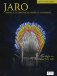
Cochlear Fluid Spaces and Structures of the Gerbil High-Frequency Region Measured Using Optical Coherence Tomography (OCT)

xlomafota13 shared this article with you from Inoreader

10162.jpg

Abstract

Since it has been difficult to directly observe the morphology of the living cochlea, our ability to infer the mechanical functioning of the living ear has been limited. Nearly all our knowledge about cochlear morphology comes from postmortem tissue that was fixed and processed using procedures that possibly distort the structures and fluid spaces of the organ of Corti. In this study, optical coherence tomography was employed to obtain volumetric images of the high-frequency hook region of the gerbil cochlea, as viewed through the round window, with far better resolution capability than had been possible before. The anatomical structures and fluid spaces of the organ of Corti were segmented and quantified in vivo and over a 90-min postmortem period. We find that the arcuate-zone and pectinate-zone widths change very little postmortem. The volume of the scala tympani between the round-window membrane and basilar membrane and the volume of the inner spiral sulcus d ecrease in the first 60-min postmortem. While textbook drawings of the mammalian organ of Corti and cortilymph prominently depict the tunnel of Corti, the outer tunnel is typically missing. This is likely because textbook drawings are typically made from images obtained by histological methods. Here, we show that the outer tunnel is nearly twice as big as the tunnel of Corti or the space of Nuel. This larger outer tunnel fluid space could have a substantial, little-appreciated effect on cochlear micromechanics. We speculate that the outer tunnel forms a resonant structure that may affect reticular-lamina motion.

View on the web

Δευτέρα 21 Φεβρουαρίου 2022

A new treatment strategy for hemifacial microsomia: Auricular reconstruction with an expanded two-flap method and simultaneous mandibular distraction osteogenesis

xlomafota13 shared this article with you from Inoreader

J Plast Reconstr Aesthet Surg. 2022 Jan 31:S1748-6815(22)00054-7. doi: 10.1016/j.bjps.2022.01.035. Online ahead of print.

ABSTRACT

BACKGROUND: Owing to the complex clinical manifestations of hemifacial microsomia (HFM), multidisciplinary cooperation is required to achieve better therapeutic effects in terms of function and aesthetics. This study aimed to explore the efficacy of the expanded two-flap auricular reconstruction combined with mandibular distraction osteogenesis in the treatment of HFM.

METHODS: This surgical strategy was performed in three stages. In the first stage, the retroauricular skin was expanded with a tissue expander and a mandibular distraction device was installed. In the second stage, the traditional expanded two-flap method for auricular reconstruction was adapted, and the framework was fabricated with costal cartilage and wrapped with the expanded skin flap, retroauricular fascia flap, and full-thickness skin graft. In the final stage, the tragus and lobule were rebuilt, the concha cavity deepened, and the mandibular distraction device removed.

RESULTS: From January 2014 to November 2018, 166 HFM patients underwent auricular reconstruction with the expanded two-flap method and simultaneous mandibular extension in our hospital. The median follow-up period was 9.3 months. Of the 166 patients, 154 patients and their families were satisfied with the results, and only 16 patients experienced complications.

CONCLUSIONS: This three-stage technique of simultaneous auricular reconstruction and mandibular distraction osteogenesis is safe and effective in achieving facial symmetry, improving occlusal contact, shortening treatment course, and relieving patient's suffering, especially for HFM patients.

PMID:35183465 | DOI:10.1016/j.bjps.2022.01.035

View on the web

Bmal1 promotes cementoblast differentiation and cementum mineralization via Wnt/β-catenin signaling

xlomafota13 shared this article with you from Inoreader
Via histochem

pubmed-meta-image.png

Acta Histochem. 2022 Feb 17;124(3):151868. doi: 10.1016/j.acthis.2022.151868. Online ahead of print.

ABSTRACT

Remodeling of the cementum plays a crucial role in periodontal regenerative therapy, while the precise mechanism of cementogenesis has yet been adequately understood. Recent studies have indicated the connection between osteogenic differentiation and Brain and muscle aryl hydrocarbon receptor nuclear translocator-like protein-1 (Bmal1). Besides, Wnt/β-catenin signal ing is proven to be an essential regulator in cementogenesis. In this study, we found a robust expression of Bmal1 in cementoblasts in the mandibular first molar of mice by immunohistochemical staining. To further explore the role of Bmal1 in cementogenesis, we examined the expression pattern of Bmal1 in OCCM-30, an immortalized murine cementoblast cell line by qRT-PCR and western blot. Our data demonstrated the upregulation of Bmal1 at both mRNA and protein levels during differentiation. Additionally, stable knockdown of Bmal1 in OCCM-30 cells resulted in downregulation of osteogenic markers such as alkaline phosphatase (Alp), osteopontin (Opn), and osteocalcin (Ocn), and reduced formation of mineralized nodules. Moreover, qRT-PCR and western blot results exhibited that the expression of β-catenin was attenuated by Bmal1 deficiency. We also found that the mRNA levels of Tcf1 and Lef1, the target transcription factors of β-catenin, were reduced by Bmal1 deficiency. In conclusion, this study preliminarily confirms that Bmal1 promotes cementoblast differentiation and cementum mineralization via Wnt/β-catenin signaling, which contributes to a potential strategy in periodontal regenerative therapy.

PMID:35183881 | DOI:10.1016/j.acthis.2022.151868

View on the web

Secondary Ewing's sarcoma of the temporo-occipital region developed 12-years after medulloblastoma treatment

xlomafota13 shared this article with you from Inoreader

Braz J Otorhinolaryngol. 2022 Jan 29:S1808-8694(22)00004-0. doi: 10.1016/j.bjorl.2022.01.003. Online ahead of print.

NO ABSTRACT

PMID:35184979 | DOI:10.1016/j.bjorl.2022.01.003

View on the web

Is there a relationship between tonsil volume and the success of pharyngeal surgery among adult patients with obstructive sleep apnea?

xlomafota13 shared this article with you from Inoreader

Braz J Otorhinolaryngol. 2022 Jan 4:S1808-8694(21)00214-7. doi: 10.1016/j.bjorl.2021.12.002. Online ahead of print.

ABSTRACT

OBJECTIVES: Hypertrophic palatine tonsils play a role in the blockage of the upper airway, one of the known causes of Obstructive Sleep Apnea (OSA). Therefore, it is possible that there is an association between tonsil size and the success of pharyngeal surgery during OSA treatment. The main objective of this study was to evaluate the relationship between tonsil grade and volume, as well as to establish whether a relationship exists between tonsil size and the success rate of pharyngeal surgery (tonsillectomy and pharyngoplasty with barbed sutures).

METHODS: This retrospective study includes forty-four adult patients who underwent tonsillectomy and pharyngeal surgery with barbed sutures for the treatment of simple snoring and OSA between January 2016 and September 2019. Patients who had been previously tons illectomized or those for whom tonsil volume measurement was lacking were excluded. All patients underwent a pre-operative physical exploration at the clinic exam room and a sleep study. Prior to surgery a Drug Induced Sleep Endoscopy (DISE) was performed. Tonsil volume was measured intraoperatively using the water displacement method. The same sleep study was repeated six months following surgery.

RESULTS: A significant correlation was found between tonsil grade and volume and between such measurements and the blockage observed at the level of the oropharynx during the DISE. Moreover, an association was observed between tonsil volume, but not tonsil grade, and the success of tonsillectomy and pharyngoplasty with barbed sutures. A tonsil volume greater than 6.5 cm3 was linked to success during pharyngeal surgery.

CONCLUSION: A correlation exists between tonsil grade and tonsil volume. A bigger tonsil volume is associated with a greater success rate of orophary ngeal surgery during treatment of OSA.

LEVEL OF EVIDENCE: Level 3, non-randomized cohort study.

PMID:35184978 | DOI:10.1016/j.bjorl.2021.12.002

View on the web

Locoregional Control and Survival in Children, Adolescents, and Young Adults With Localized Head and Neck Alveolar Rhabdomyosarcoma-The French Experience

xlomafota13 shared this article with you from Inoreader

Front Pediatr. 2022 Feb 4;9:783754. doi: 10.3389/fped.2021.783754. eCollection 2021.

ABSTRACT

INTRODUCTION: The head and neck (HN) are the most frequent sites of pediatric rhabdomyosarcoma (RMS). Alveolar RMS (ARMS) represents ~20% of all RMS cases and frequently spread to lymph nodes (LNs). The aim was to report locoregional control, event-free survival (EFS), and overall survival (OS), according to clinical and pathological features, LN staging, and treatment modalities.

METHODS: The study included all patients prospectively enrolled in EpSSG RMS 2005 study under 21 years of age with localized HN ARMS and diagnosed between 2005 and 2016 in France. Medical data including imaging, surgical report, and radiation therapy planes were analyzed.

RESULTS: Forty-eight patients (median age 6 years; range 4 months-21 years), corresponding to 30 parameningeal and 18 non-parameningeal ARMS, were included. There were 33 boys (69%). Tumor loca tions included the following: orbit (n = 7) among which four cases had bone erosion, paranasal sinuses and nasal cavity (n = 16), deep facial spaces (n = 10), nasolabial fold (n = 8), and other non-parameningeal HN sites (n = 7). A fusion transcript of PAX3-FOXO1 or PAX7-FOXO1 was expressed in 33 of the 45 cases (73%) with molecular analysis. At diagnosis, 10 patients had primary resection of the primary tumor (PRPT) (none with microscopic complete resection) and 9 had LN staging. After induction chemotherapy, 26 patients (54%) had secondary resection of the primary tumor (SRPT) and 13 patients (27%) had cervical LN dissection. A total of 43 patients (90%) were treated with radiation therapy.With a median follow-up of 7 years (range 2-13 years), 5-year OS and EFS were 78% (95% CI, 63-88%) and 66% (95% CI, 51-78%), respectively. We observed 16 events (10 deaths): 4 local, 4 regional, 1 local and regional, and 7 metastatic. In univariate analysis, OS was only superior for patients under 10 years of age (p = 0.002), while FOXO1-negative ARMS, SRPT for parameningeal ARMS, and LN surgery were associated with significantly better EFS.

CONCLUSION: Our study confirms a better outcome for fusion-negative ARMS and ARMS in children under 10 years. Moreover, LN surgery and SRPT of parameningeal tumor may improve EFS of ARMS. Larger studies are needed to confirm our findings.

PMID:35186818 | PMC:PMC8855824 | DOI:10.3389/fped.2021.783754

View on the web

Analysis of effects of laryngopharyngeal reflux disease and proton pump inhibitor treatment on Eustachian tube function in patients with obstructive sleep apnea hypopnea

xlomafota13 shared this article with you from Inoreader

pubmed-meta-image.png

Ear Nose Throat J. 2022 Feb 21:1455613221076786. doi: 10.1177/01455613221076786. Online ahead of print.

ABSTRACT

OBJECTIVE: This study aims to explore the effects of laryngopharyngeal reflux disease (LPRD) and proton pump inhibitor (PPI) treatment on Eustachian tube function in patients with obstructive sleep apnea (OSA).

METHODS: The Eustachian tube score-7 (ETS-7) was observed before and after PPI treatment in the control group, OSA only group, and OSA + LPRD group.< /p>

RESULTS: Age, sex, smoking history, and drinking history showed no differences among 3 groups (P > .05). The body mass index (BMI) in the control group was lower than that in other groups (P < .017). Before PPI treatment, the abnormality rate of ETS-7 in the OSA + LPRD group statistically differed from that in the control group and the OSA only group (P < .017). After PPI treatment, the abnormality rate of ETS-7 in the OSA + LPRD group exhibited no significant differences compared with that in the control group and the OSA only group (P > .017), and it declined remarkably compared with that before PPI treatment (75% vs 35%, χ2 = 13.334, P = .001). Moreover, the multivariate analysis revealed that only LPRD had an independent correlation with the abnormality of ETS-7 (OR = 1.245, 95% CI: 1.759-6.861, P = .000).

CONCLUSION: In view of its high incidence in OSA patients, LPRD may be a considerable factor for the high incidence of abnormality r ate of ETS-7 in OSA patients, and PPI therapy is of significant value in improving Eustachian tube function in OSA patients with LPRD.

PMID:35187968 | DOI:10.1177/01455613221076786

View on the web

Αρχειοθήκη ιστολογίου