Παρασκευή 10 Απριλίου 2020

Paediatric Day Case Tonsillectomy - Audit of a New Programme.

Paediatric Day Case Tonsillectomy - Audit of a New Programme.:

Related Articles
Paediatric Day Case Tonsillectomy - Audit of a New Programme.

Ir Med J. 2020 Apr 03;113(4):56

Authors: Grant O, Harper P

Abstract

Aims The primary objective of this audit was to assess 30-day unplanned admission or readmission rates following day case tonsillectomy. Secondary objectives included assessing demographic characteristics of patients and surgical and anaesthetic techniques employed. Methods Retrospective chart review was performed. Results 34 tonsillectomies or adenotonsillectomies were performed for children aged between 5 and 17 years. A total of six patients (17.6%) were either admitted from the day ward or readmitted within 30 days. This was not statistically significantly greater than the maximum acceptable rate of 15% as recommended by ENT UK and the Royal College of Surgeons (95% confidence intervals 4.8% - 30.5%, p = 0.33). Of these admissions, one (2.9%) was due to nausea and inadequate oral intake, one (2.9%) was due to clinical concerns regarding sleep apnoea and four readmissions (11.8%) were due to bleeding. Anaesthesia techniques used varied considerably. Conclusion Further multidisciplinary co-operation and standardisation of care may help to improve this programme and reduce unplanned admission and readmission rates.

PMID: 32268049 [PubMed - as supplied by publisher]

COVID‐19 pandemic: effects and evidence‐based recommendations for otolaryngology and head and neck surgery practice

COVID‐19 pandemic: effects and evidence‐based recommendations for otolaryngology and head and neck surgery practice:

hed.v42.4.cover.gif

Abstract

The 2019 novel coronavirus disease (COVID‐19) is a highly contagious zoonosis produced by SARS‐CoV‐2 that is spread human‐to‐human by respiratory secretions. It was declared by the WHO as a public health emergency. The most susceptible populations, needing mechanical ventilation, are the elderly and people with associated comorbidities.

There is an important risk of contagion for anesthetists, dentists, head and neck surgeons, maxillofacial surgeons, ophthalmologists and otolaryngologists. Health workers represent between 3.8% to 20% of the infected population; some 15% will develop severe complaints and among them, many will lose their lives. A large number of patients do not have overt signs and symptoms (fever/respiratory), yet pose a real risk to surgeons (who should know this fact and must therefore apply respiratory protective strategies for all patients they encounter).

All interventions that have the potential to aerosolize aerodigestive secretions should be avoided or used only when mandatory. Health workers who are: pregnant, over 55‐65 years of age, with a history of chronic diseases (uncontrolled hypertension, diabetes mellitus, chronic obstructive pulmonary diseases and all clinical scenarios where immunosuppression is feasible, including that induced to treat chronic inflammatory conditions and organ transplants) should avoid the clinical attention of a potentially infected patient. Healthcare facilities should prioritize urgent and emergency visits and procedures until the present condition stabilizes; truly elective care should cease and discussed on a case‐by‐case basis for cancer patients.

For those who are working with COVID‐19 infected patients' isolation is compulsory in the following settings: a) unprotected close contact with COVID‐19 pneumonia patients: b) onset of fever, cough, shortness of breath and other symptoms (gastrointestinal complaints, anosmia and dysgeusia have been reported in a minority of cases).

For any care or intervention in the upper aerodigestive tract region, irrespective of the setting and a confirmed diagnosis (eg, ; rhinoscopy or flexible laryngoscopy in the outpatient setting and tracheostomy or rigid endoscopy under anesthesia) it is strongly recommended that all healthcare personnel wear personal protective equipment (PPE) such as N95, gown, cap, eye protection and gloves.

The procedures described are essential in trying to maintain safety of healthcare workers during COVID‐19 pandemic. In particular, otolaryngologists, head and neck, and maxillofacial surgeons are per se exposed to the greatest risk of infection while caring for COVID‐19 positive subjects, and their protection should be considered a priority in the present circumstances.

This article is protected by copyright. All rights reserved.

Ceftriaxone‐induced radiation recall dermatitis

Ceftriaxone‐induced radiation recall dermatitis:

hed.v42.4.cover.gif

Abstract

Background

Radiation recall dermatitis (RRD) is an acute inflammatory skin reaction occurring in a skin area previously exposed to radiotherapy and triggered by subsequent intake of a drug, most commonly a chemotherapeutic agent. RRD secondary to antibiotics has also been reported but is a rare phenomenon overall and there are no reports of RRD in association with ceftriaxone exposure.

Methods

We report on a 59‐year‐old patient who had received radiotherapy to the neck bilaterally and who developed RRD 6 months later after a single dose of intramuscular ceftriaxone.

Results

The patient's rash resolved without further intervention over the ensuing 2 days following administration of a single dose of ceftriaxone.

Conclusion

This case illustrates that while RRD secondary to antibiotic exposure is rare, it is part of the differential diagnosis to be considered for acute dermatitis when there is a past history of radiotherapy to the same skin area.

Multiple metabolic parameters and visual assessment of 18 F-FDG uptake heterogeneity of PET/CT in advanced gastric cancer and primary gastric lymphoma

Multiple metabolic parameters and visual assessment of 18 F-FDG uptake heterogeneity of PET/CT in advanced gastric cancer and primary gastric lymphoma:

Abstract


Purpose

Advanced gastric cancer (AGC) and primary gastric lymphoma (PGL) are the two most common malignant tumors of the stomach. Conventional imaging examinations have difficulty distinguishing the two. This study explored the values of multiple parameters and visual assessment of 18F-fluorodeoxyglucose(18F-FDG) uptake heterogeneity of positron emission tomography/computed tomography(PET/CT) for differentiating between AGC and PGL.


Methods

This retrospective study included 70 AGC and 26 PGL patients, all of whom had undergone 18F-FDG PET/CT before treatment. We analyzed the differences between AGC and PGL in the distribution of metastatic lesions and multiple metabolic parameters, including the maximum standardized uptake value (SUVmax), SUVmax/maximal thickness(THKmax), metabolic tumor volume and total lesion glycolysis (TLG). In addition, 18F-FDG uptake heterogeneity was visually assessed using a visual scoring method and a method of measuring SUVmax differences (SUVmax-d).


Results

The most common metastasis of AGC patients were liver, bone, peritoneal and proximal lymph nodes; PGL patients had fewer peritoneal metastases and lymph node metastasis could appeared to be “skip metastasis.” The metabolic parameters—SUVmax, SUVmax/THKmax and TLG—were higher in patients who had PGL, especially in diffuse large B-cell lymphoma (DLBCL). In the visual assessment of 18F-FDG uptake heterogeneity, the measurements of SUVmax-d in PGL were significantly higher than in AGC. Receiver operating characteristics curve analysis suggested that SUVmax has the highest comprehensive diagnostic efficiency due to having the highest value of area under the curve and the highest accuracy (77.2%).


Conclusion

18F-FDG PET/CT had a high diagnostic efficiency for discrimination of AGC and PGL, especially between DLBCL and other pathological subtypes. Visual assessment used to evaluate 18F-FDG uptake heterogeneity could help to distinguish the two types of tumors. In addition, our innovative method of measuring the heterogeneity of 18F-FDG uptake—namely, SUVmax-d—could contribute to identification of the two tumor types and should have its significance clarified by future studies.

Cancers, Vol. 12, Pages 919: Artesunate Affects T Antigen Expression and Survival of Virus-Positive Merkel Cell Carcinoma

Cancers, Vol. 12, Pages 919: Artesunate Affects T Antigen Expression and Survival of Virus-Positive Merkel Cell Carcinoma:

cancers-12-00919-g001-550.jpg


Cancers, Vol. 12, Pages 919: Artesunate Affects T Antigen Expression and Survival of Virus-Positive Merkel Cell Carcinoma

Cancers doi: 10.3390/cancers12040919

Authors:
Bhavishya Sarma
Christoph Willmes
Laura Angerer
Christian Adam
Jürgen C. Becker
Thibault Kervarrec
David Schrama
Roland Houben


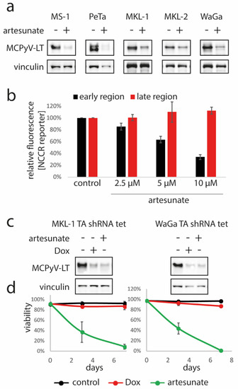
Merkel cell carcinoma (MCC) is a rare and highly aggressive skin cancer with frequent viral etiology. Indeed, in about 80% of cases, there is an association with Merkel cell polyomavirus (MCPyV); the expression of viral T antigens is crucial for growth of virus-positive tumor cells. Since artesunate—a drug used to treat malaria—has been reported to possess additional anti-tumor as well as anti-viral activity, we sought to evaluate pre-clinically the effect of artesunate on MCC. We found that artesunate repressed growth and survival of MCPyV-positive MCC cells in vitro. This effect was accompanied by reduced large T antigen (LT) expression. Notably, however, it was even more efficient than shRNA-mediated downregulation of LT expression. Interestingly, in one MCC cell line (WaGa), T antigen knockdown rendered cells less sensitive to artesunate, while for two other MCC cell lines, we could not substantiate such a relation. Mechanistically, artesunate predominantly induces ferroptosis in MCPyV-positive MCC cells since known ferroptosis-inhibitors like DFO, BAF-A1, Fer-1 and β-mercaptoethanol reduced artesunate-induced death. Finally, application of artesunate in xenotransplanted mice demonstrated that growth of established MCC tumors can be significantly suppressed in vivo. In conclusion, our results revealed a highly anti-proliferative effect of the approved and generally well-tolerated anti-malaria compound artesunate on MCPyV-positive MCC cells, suggesting its potential usage for MCC therapy.

Clinical Practice Guideline: Ménière's Disease.

Clinical Practice Guideline: Ménière's Disease.:

pubmed256blue.png


Related Articles
Clinical Practice Guideline: Ménière's Disease.

Otolaryngol Head Neck Surg. 2020 Apr;162(2_suppl):S1-S55

Authors: Basura GJ, Adams ME, Monfared A, Schwartz SR, Antonelli PJ, Burkard R, Bush ML, Bykowski J, Colandrea M, Derebery J, Kelly EA, Kerber KA, Koopman CF, Kuch AA, Marcolini E, McKinnon BJ, Ruckenstein MJ, Valenzuela CV, Vosooney A, Walsh SA, Nnacheta LC, Dhepyasuwan N, Buchanan EM

Abstract

OBJECTIVE: Ménière's disease (MD) is a clinical condition defined by spontaneous vertigo attacks (each lasting 20 minutes to 12 hours) with documented low- to midfrequency sensorineural hearing loss in the affected ear before, during, or after one of the episodes of vertigo. It also presents with fluctuating aural symptoms (hearing loss, tinnitus, or ear fullness) in the affected ear. The underlying etiology of MD is not completely clear, yet it has been associated with inner ear fluid (endolymph) volume increases, culminating in episodic ear symptoms (vertigo, fluctuating hearing loss, tinnitus, and aural fullness). Physical examination findings are often unremarkable, and audiometric testing may or may not show low- to midfrequency sensorineural hearing loss. Conventional imaging, if performed, is also typically normal. The goals of MD treatment are to prevent or reduce vertigo severity and frequency; relieve or prevent hearing loss, tinnitus, and aural fullness; and improve quality of life. Treatment approaches to MD are many and typically include modifications of lifestyle factors (eg, diet) and medical, surgical, or a combination of therapies.

PURPOSE: The primary purpose of this clinical practice guideline is to improve the quality of the diagnostic workup and treatment outcomes of MD. To achieve this purpose, the goals of this guideline are to use the best available published scientific and/or clinical evidence to enhance diagnostic accuracy and appropriate therapeutic interventions (medical and surgical) while reducing unindicated diagnostic testing and/or imaging.

PMID: 32267799 [PubMed - as supplied by publisher]
Plain Language Summary: Ménière's Disease.

Plain Language Summary: Ménière's Disease.:

pubmed256blue.png


Related Articles
Plain Language Summary: Ménière's Disease.

Otolaryngol Head Neck Surg. 2020 Apr;162(4):435-445

Authors: Basura GJ, Colandrea M, Walsh SA, Kuch AA, Monjur TM

Abstract

This plain language summary explains Ménière's (pronounced men-yerz) disease (MD) to patients. The summary applies to patients aged 18 years and older with a suspected diagnosis of definite or probable MD. It is based on the 2020 "Clinical Practice Guideline: Ménière's Disease." This guideline uses published research to best advise health care providers and patients on the history and physical examination of patients with MD and how to diagnose and treat them. The guideline includes recommendations that are explained in this summary. Recommendations may not apply to every patient but can be used to facilitate shared decision making between patients and their health care providers.

PMID: 32267819 [PubMed - as supplied by publisher]

Αρχειοθήκη ιστολογίου