Cancers, Vol. 12, Pages 1074: Imaging Accuracy in Preoperative Staging of T3-T4 Laryngeal Cancers
Cancers doi: 10.3390/cancers12051074
Authors:
Marco Benazzo
Fabio Sovardi
Lorenzo Preda
Simone Mauramati
Sergio Carnevale
Giulia Bertino
Francesca Berton
Matteo Meroni
Irene Herman
Giuseppe Trisolini
Patrizia Morbini
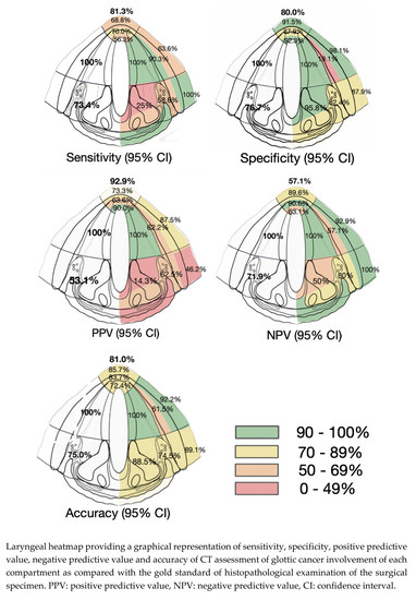
Background: Preoperative imaging impacts treatment planning and prognosis in laryngeal cancers. We investigated the accuracy of standard computed tomography (CT) in evaluating tumor invasions at critical glottic areas. Methods: CT scans of glottic cancers treated by partial or total laryngectomy between Jan 2015 and Aug 2019 were reviewed to assess levels of tumor invasion at critical glottic subsites. CT accuracy in the identification of tumor extensions was determined against the gold standard of histopathological analysis of surgical samples. Results: This study included 64 patients. In the anterior commissure, CT showed high rates of false positives at all levels (sensitivity 56.2–70%, specificity 87.8–92.3%); in the anterior vocal fold, it overestimated the deep invasion (19.5% specificity, 90.3% sensitivity), while it underestimated the extralaryngeal spread (63.6% sensitivity, 98.1% specificity). In the posterior paraglottic space (pPGS), false negative results were more frequent for superficial extensions (25% sensitivity, 95.8% specificity) and deep invasions (58.8% sensitivity, 82.3% specificity). Shorter disease-specific and disease-free survivals were associated with pStage IV (p: 0.045 and 0.008) and with the pathological involvement of pPGS (p: 0.045 and 0.015). Conclusions: Negative prognostic correlation of pPGS involvement was confirmed on histopathological data. CT staging did not provide a satisfactory prognostic stratification and should be complemented with magnetic resonance imaging.

Δεν υπάρχουν σχόλια:
Δημοσίευση σχολίου